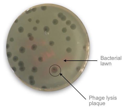
The growing number of antibiotic-resistant bacterial infections (antibiotic resistance), a consequence of the overuse of antibiotics, has been identified by the WHO as one of the main public health threats by 2050. Phage therapy, based on the use of bacterial viruses (bacteriophages), is generating considerable interest in this context. However, in order to be implemented, it requires the identification of the active phage on a given bacterium in order to destroy it. This assessment is currently based on visual detection of lysis plaques formed by bacteriophages on Petri dishes covered with bacteria (
Figure). This visual inspection, both manual and excessively time-consuming (12-24h), limits the possibilities of applying phage therapy to the patient.
 Phage lysis plaque.
 To test the sensitivity of a bacterium to a bacteriophage, the two organisms are co-cultured on the surface of an agar-coated Petri dish where a bacterial lawn forms. If the bacterium is sensitive to the bacteriophage, it is killed (lysis) and a phage lysis plaque is formed on the surface of the agar plate as the lysis progresses.
© Pheliqs
An original approach has been proposed by researchers from our laboratory, the Department of Technologies for Biology and Health (DTBS), the Microelectronics Technology Laboratory, and Swiss researchers [a
collaboration between photonics researchers, biophysicists and microbiologists] to develop and test optical systems to verify phage-bacteria matching very quickly. The chosen strategy is to develop a lensless imaging system around a large format CMOS optical sensor (
Figure). The analysis of lysis plaques is based on the use of an algorithm developed in-house, which allows to reconstruct the images diffracted by the bacteria in order to determine in real time the number and the kinetics of the lysis plaques. These measurements provide information on the nature and efficiency of phages against bacteria and allow to determine their titer, to study their morphologies and their growth kinetics. This new lens-free imaging approach has several advantages. Firstly, in the absence of lenses, the resolution and the field of view of the image are only limited by the pixel pitch and the size of the sensor. However, the current technologies drawn by the digital imaging sector allow the realization of very large sensors with very small pixels (typically several tens of million pixels on a sensor of 24 by 36 mm). Consequently, a much larger field of view than that of conventional optical microscopy is available for monitoring phage-bacteria interaction. Moreover, since the pixel pitch of current sensors is only a few microns, lens-free imaging allows to resolve structures of a few tens of microns, such as nascent bacterial micro-colonies, and thus to save time in the analysis process.
 Custom prototype without lens.
 The device is composed of a detection system and an illumination module. The sensing system consists of a 22.3 × 14.9 mm
2 APS-C (Advanced Photo System type-C) CMOS (Complementary Metal Oxide Semiconductor) sensor from a "consumer" digital camera. The sensor consists of a 5344 × 3516 pixel matrix with a pitch of 4.3 μm. The Petri dish on which the bacteriophages are located is placed directly on the sensor and illuminated from above by a monochromatic green LED (518 nm) coupled to a 200 μm diameter multimode optical fiber (Thorlabs M72L02). The images are acquired directly and converted to .tiff files. They are then processed using two different algorithms; the first processes the entire image area to detect plaques, while the second processes only a cropped subimage of each plaque to calculate the growth rate.
© Pheliqs
Using this technique, the researchers report that they determined the susceptibility of
Staphylococcus aureus to different phage after only 3 hours and the infectious titer after 8 hours and 20 minutes. These times are much shorter than the 12 to 24 hours usually required for naked-eye observation and counting of lysis plaques. In addition, continuous monitoring of the samples allowed the study of plate growth kinetics and confirmation of the correlation between bacterial density and phage diffusion in the agar layer. Finally, with the resolution of 4.3 μm (
Figure), the researchers were able to detect phage-resistant Klebsiella pneumoniae bacterial microcolonies within the boundaries of the lysis plates, showing that their prototype is also a suitable device for monitoring phage resistance.
This first proof of concept of a lens-free imaging system, implemented as a compact and economical device, opens a promising avenue for phage therapy. Several national and international programs are underway to validate its applications. Future work will also focus on the development of more refined algorithms allowing the morphological classification of plaques (and therefore phages) according to their growth rate but also their morphotype.
Collaboration: Quantum Photonics, Electronics and Engineering (IRIG, CEA-Grenoble, France); Department of Microtechnologies for Biology and Health (Leti-DTBS, CEA-Grenoble, France); LTM–Micro and Nanotechnologies for Health (CNRS, France); Department of Fundamental Microbiology (University of Lausanne, Switzerland).