The distribution of the electromagnetic field is an essential input for the design and development of new photonic structures. The classical near-field microscopy tools (SNOM, PSTM ..) allow to obtain this information experimentally. This type of scanning probe microscopy is mainly based on a point by point record of the evanescent field obtained through a tapered fiber who sweeps the surface under investigation at a few tens of nanometer distance. These techniques require both complex equipment but also a highly skilled experimentalist.
We have developed an original approach, simple and inexpensive, to obtain electromagnetic field maps with substantially the same resolution as that obtained by near field microscopy. This approach consists in studying with a standard optical microscopy the trajectory of fluorescent beads (500 nm) moving in the near field of the optical component.
This innovative optofluidic approach that combines colloids and photonics resolves the spatial distribution of the electromagnetic field with a resolution 10 times smaller than the wavelength.
Experimentally, the photonic structure (here an optical microcavity) to be investigated is placed in a microfluidic cell filled with a colloidal suspension. The colloidal particles are subjected to Brownian motion and follow random trajectories. Some of them will "fall" into the optical trap generated by the microcavity. Within this trap, the flow of particles dictated by optical forces reveals the field map of the resonant modes.
This original approach developed in close collaboration with the “Micronanotechnology for health” team at LTM and the Field Optics group of Dijon (Sinoptiq LRC) opens promising prospects in biotechnology, such as the study and manipulation of biological objects (bacteria, cells).
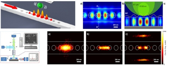
Figure: (Top) Schematic of the SOI microcavity and modeling of the electromagnetic field. (Down) Experimental set-up and electromagnetic field experimental distribution as mapped using a 2µm (a), 1 µm (b) or 500nm (c) diameter fluorescent bead.